


What is craniopharyngioma?
This type of benign tumour or cystic mass is quite rare and congenital (from birth). 50% occur in children under 16 years, and the remainder at any time in adult life. The tumour exerts pressure on the hypothalamus which lies just above the pituitary gland and is responsible for releasing hormones that start and stop the release of pituitary hormones.
Faster growing craniopharyngiomas affect children whilst slower growing ones affect adults. This type of tumour can cause headaches and vision problems and can affect hunger, thirst and sleep patterns.
The onset of puberty and growth hormone production in children can also be affected, plus growth hormone production may be affected in adults.
The tumours can be solid, cystic (full of fluid), calcified, or full of debris. They are slow-growing tumours that can take 2-3 years (or longer) to manifest themselves before a diagnosis is made.
Symptoms
There are a range of symptoms that affect people with craniopharyngioma, you may not experience all of these:
- headaches (sometimes accompanied by nausea or vomiting)
- AVPDeficiency(Diabetes Insipidus)
- disturbed sleep patterns
- vision disturbance
- behavioural changes, including introversion and inability to concentrate
- slow growth
- increased sensitivity to cold or heat
- early or delayed puberty -children
- irregular periods or loss of normal menstrual function (Amenorrhoea) -adult females
- impotence -adult males
- reduced fertility -adults
- decrease in sex drive -adults
- tiredness and susceptibility to infections
- appetite and weight variations
Diagnosis
Investigations will be carried out to find out as much as possible about the type, position and size of the tumour. This will help them decide on the best treatment option. These tests include:
- CT scan
- MRI scan
- ophthalmic assessment
- endocrine assessment of blood hormone levels
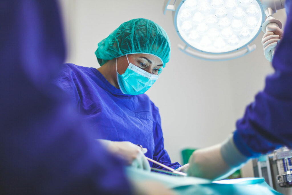
Treatment
Surgery is the main treatment strategy for craniopharyngioma, and if possible, the neurosurgeon will try to remove the tumour.However, surgery can cause some damage to the surrounding structures, and often the tumour will be left behind intentionally to minimise this risk. Radiotherapy may then be given to stop the tumour from growing. After surgery further, further tests will be done to check hormone levels. If hormones are depleted then replacement therapies will be given. A follow up MRI scan may be necessary to ensure stability of the tumour especially if removal was not possible.