




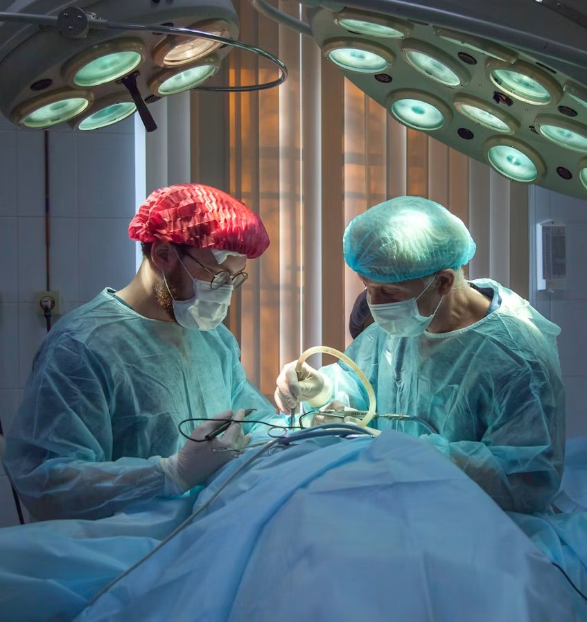
We were very pleased to see pituitary news make headlines recently, with the world’s first artificial intelligence-assisted surgery taking place on a pituitary tumour. Rhys Hibbert, a volunteer of The Pituitary Foundation, was the first person in the world to undergo AI-assisted pituitary surgery earlier this year.
Rhys, a father of two from Bedfordshire, underwent transsphenoidal pituitary surgery earlier this year. Rhys was diagnosed with an 11mm pituitary tumour after he fell unconscious and had a seizure during a morning walk. Rhys also noticed loss of his peripheral vision, as well as dizziness and fatigue.
As the pituitary gland is very close to the optic nerves, it is common that tumours on the gland can press on the nerve as they grow. This can cause changes to vision, like blurred vision, double vision or reduced peripheral vision. For some people, vision changes may be the first symptom they experience of a tumour.
The surgery was carried out by Professor Hani Marcus and Mr Danyal Khan, at the National Hospital for Neurology and Neurosurgery. Hani is a pituitary neurosurgeon and a member of our Board of Trustees, and is very committed to supporting the pituitary community both through his clinical work and his research interests. In addition to his work utilising AI in surgery, he has also been involved in research into the development of robotics-assisted surgery. You can read more about this here.
What does AI-assisted surgery involve?
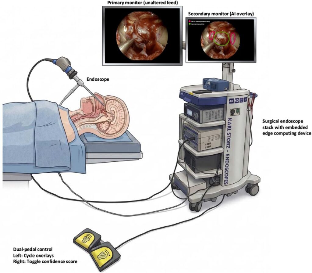
Traditionally, operations on the pituitary gland are carried out using a technique called transsphenoidal surgery. ‘Trans’ means across and ‘sphenoid’ is the air cavity in which the pituitary gland sits. Transsphenoidal surgery involves going through the nose to access the skull and feeding a small camera (an endoscope) through the nose so that surgeons can watch a video stream of the surgery and see what cuts to make.
Where the AI plays a part is in analysing the live-stream video feed of the surgery. This type of technology is called computer vision AI (CVAI). The CVAI shows on the video feed where blood vessels, nerves and other tissue are, to help surgeons avoid damaging them during surgery. It also helps identify where the safest places are to make cuts and remove the tumour.
Why was AI used for pituitary surgery?
The pituitary gland sits very close to important nerves and blood vessels, including the optic nerves (that control vision) and the carotid arteries (which control blood flow to the brain). This means that performing surgery exactly, to avoid damaging these nerves and blood vessels is crucial.
In addition to this, as pituitary tumours are so rare, even the most experienced pituitary surgeons will only carry out a relatively small number of pituitary surgeries in their career. Most surgeons perform, on average, 10 to 20 pituitary surgeries per year.
This presents a great opportunity for AI technology to provide a lot of benefit. Pituitary surgery carries a high risk, given how intricate and complex it is and the potential for damage to important blood vessels and nerves. Technology that can improve accuracy in these procedures could be incredibly valuable for patients.
Furthermore, the AI model used for this surgery was trained on hundreds of videos of AI surgeries, which is far more than any surgeon would be able to see and experience. This experience means the AI model is very good at being able to recognise hidden anatomical structures and highlight these in real time to surgeons carrying out the operation. This will hopefully reduce the chances of complications following the surgery, such as incomplete removal of the tumour or accidental damage to blood vessels or nerves.
Is AI-assisted surgery safe, and how could it help patients in the future?
Before reaching patients, the UCL team carried out several years of development and testing. This included identifying which anatomical structures are most important for AI to recognise, how accurately their outlines could be traced, when that information should be displayed during surgery, and how best to communicate the model’s confidence to the surgeon. The system was also tested in digital and high fidelity surgical simulations before being used in patients.
The first clinical study was deliberately small and designed primarily to test whether the technology could be used safely and practically in the operating theatre. The AI display is separate from the normal surgical video, is switched off by default, and the surgeon controls when it appears using a foot pedal. The system also displays how confident it is in its prediction, allowing the surgeon to make well-informed decisions on how best to use the information the model displays.
In the first six operations in which the system was successfully used, there were no AI-related complications, no significant distraction to the surgeon and no disruption to the operating theatre team. However, this is still very early research and the study was not designed to show that AI reduces complications. Larger studies are now needed to establish whether it can ultimately improve outcomes. This will be the real test for the model, and will be where the real benefit to patients can be shown.
The hope is that more reliable identification of structures such as the carotid arteries and pituitary region could eventually help surgeons operate more safely. In principle, that might reduce complications including vascular or neurological injury and perhaps help preserve normal pituitary function, but we do not yet have evidence that it does so. In this early study, hormone outcomes and length of hospital stay were broadly similar to previous patients undergoing conventional surgery.
What does the future of AI-assisted surgery look like?
This technology is still at an early stage. The next step is a larger study at Queen Square involving more surgical teams, followed by evaluation across multiple hospitals. Patients specifically consent to take part in the research, and the surgeon remains in control throughout, including deciding when to display the AI guidance.
Over time, we hope these systems will combine the live surgical video with other information, such as pre-operative scans and navigation systems, to give surgeons a richer picture of the anatomy in front of them. The same principle could potentially be applied to other complex operations where recognising and protecting critical structures is important.
The long-term vision is for AI to act almost like another expert in the operating theatre: a second pair of eyes that is constantly looking at the surgical field and can highlight important anatomy or potential areas of concern. It would not replace the surgeon or make decisions independently, but provide additional information to support judgement at key moments during an operation.
This article was written by Amy Shingler, Information and Communications Manager at The Pituitary Foundation, and Prof Hani Marcus, Consultant Neurosurgeon and trustee with The Pituitary Foundation.
It was written in September 2026.
If you would like to know more about this work, you can read the full research article from the UCL team.
Help support our work!
If you found this article useful, please consider donating to allow us to continue producing articles like this.