



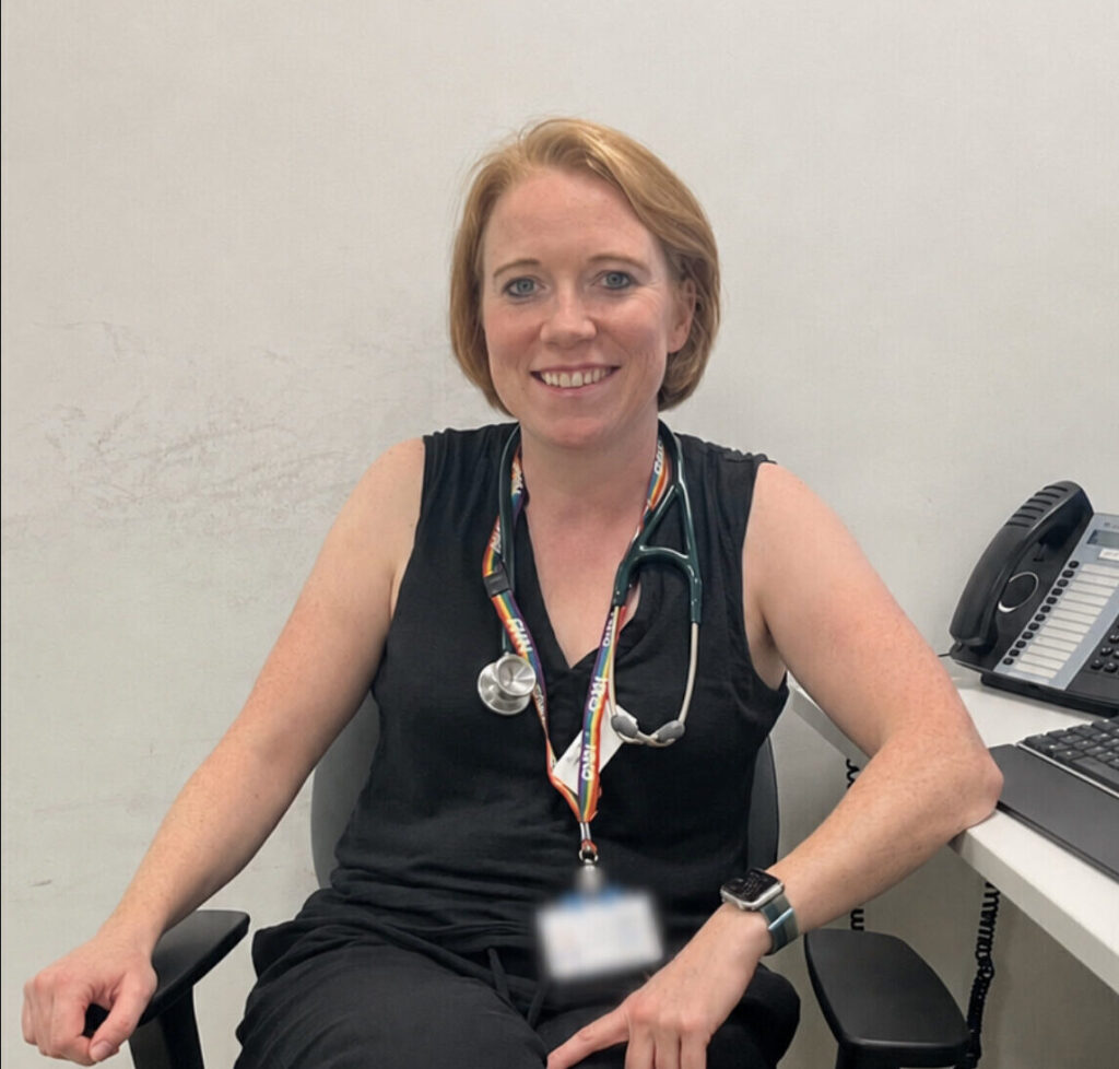
My name is Helen, I am 44 years old and an advanced clinical practitioner in an acute London hospital. On December 28, 2025, after a wonderful Christmas with the family, I suddenly developed blurred vision in my left eye from the peripheral inwards. I only noticed after covering my right eye and looking just through the left. I realised I couldn’t see about 75% of the texts on my phone reading from left to right. Everything was distorted and pictures on the walls and the clock face were only half visible.
I went to the local hospital A and E (where I also worked) and told them I thought I had a detached retina. The doctors agreed and told me to report at the eye casualty department in another hospital for 9am the following day.
“I realised I couldn’t see about 75% of the texts on my phone reading from left to right. Everything was distorted and pictures on the walls and the clock face were only half visible.”
After a sleepless night, I went to eye casualty where the ophthalmologist had a good look at the back of both eyes and gave me an OCT, a scan that looks at the retinas. They found that the jelly of the eye had come off the retina in my left eye – a ‘posterior vitreous detachment (PVD)’ that can happen with age and in people who are very short sighted like me. They said it was fine, a natural ageing process and the retinas were intact. I would have flashes and floaters but would get used to that and eventually not notice it as much. They also did visual field tests which were fine, so they discharged me. I did say to the discharging doctor, isn’t it weird that when I cover my right eye, I can only see half your face?
For the next week I went to work, still very anxious and unable to see well. My family kept telling me to keep busy and distract myself, with my then partner saying, “this is the new normal.” But I was not reassured. How could an otherwise fit and well 44-year-old lose 75% of vision and it be normal? By this point the peripheral vision in my right eye was also starting to blur. I work in a medical team, and my team were very concerned about my symptoms. I started to read up and look at what flashes and floaters look like, realising these were not my symptoms. My colleagues begged me to go back to eye casualty the next day and not come to work. Something was just not right.
Luckily my sister was able to come with me this time. Armed with a list of questions, we saw the doctor I’d seen before. Again, a whole battery of tests proved normal. It was very useful to have a third party there. My sister was adamant, “Helen does not have the flashes and floaters that you associate with PVD, she describes it to me as looking through frosted glass”. The doctor then said, “well it might not be PVD, if that reassures you.” I was not reassured since earlier they had discharged me with that diagnosis. However, they offered to scan my head (CT), so I had to go back to A and E.
The CT found a large 3.5-centimetre lesion in my brain that was pushing my optic nerves apart and attached to my pituitary gland. It was so big it had pushed one of my optic nerves onto an internal carotid artery. I needed a contrast MRI asap.
My sister was in complete shock when we were told, I remember her hands shaking as she put down her cup of tea. She says she remembers I went completely calm, she says she still can’t describe it. My first thought was, well at least I know what’s causing my vision problem, and I haven’t been wrong to trust my gut and insist on further investigation. Had I caused this by not living as healthy as I could, I wondered? My sister put that thought straight out of my head, reassuring me this was not my fault.
“My first thought was, well at least I know what’s causing my vision problem, and I haven’t been wrong to trust my gut and insist on further investigation.”
It took an agonising week to get the MRI which revealed a possible craniopharyngioma, amongst other benign differentials. I hugged the doctor who told me as none of the differentials were cancerous, I felt so relieved. Little did I know how dangerous a craniopharyngioma could be. As they say, benign is not fine.
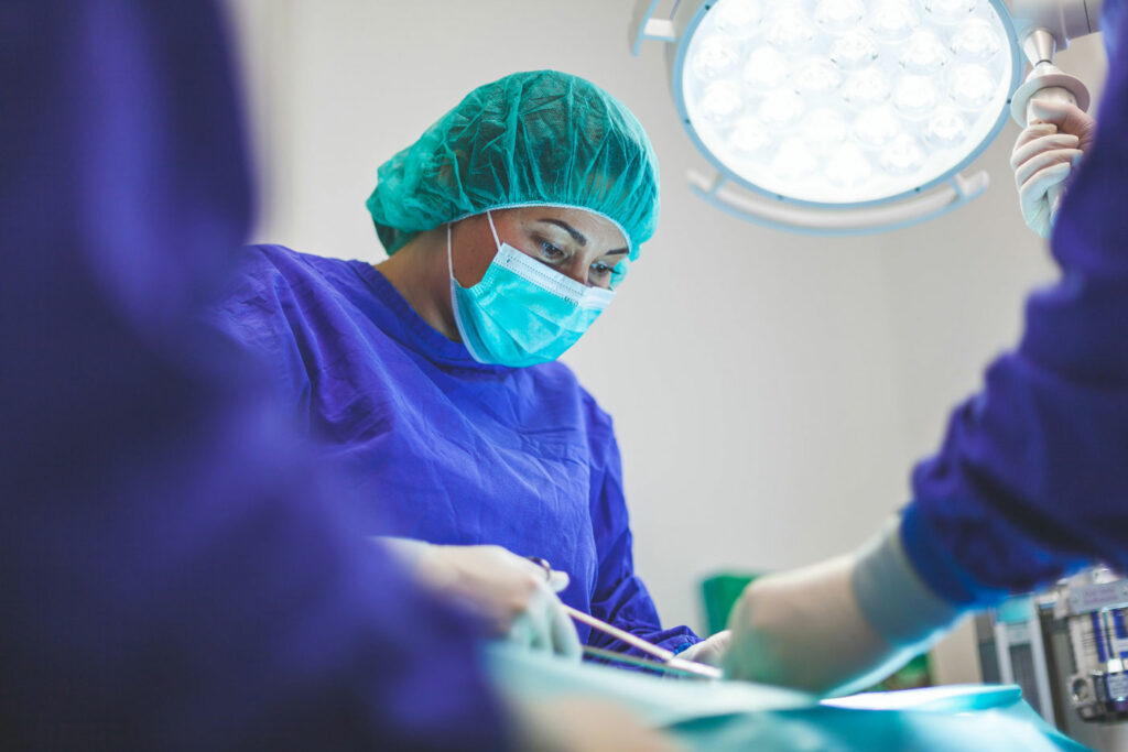
I had a craniotomy on the 13th of March 2026. The surgery took about nine hours and was a big success. The surgeon managed to remove all of the tumour and said he could not have gone any further without harming me. I awoke from surgery and the first thing I did was check my vision. To my delight and thanks to the skill of the surgeon, I could see again! It was the best feeling in the world!
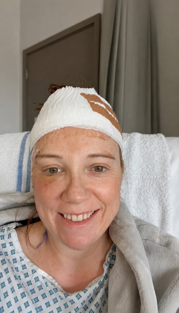
“To my delight and thanks to the skill of the surgeon, I could see again! It was the best feeling in the world!”
Histology later verified this was indeed an adamantinomatous craniopharyngioma (ACP). Post operatively it was not as plain sailing. Because of the attachment to the pituitary gland my sodium level dropped to dangerously low levels and the endocrine team struggled to control it.
I spent three weeks in high dependency and finally my sodium was brought under control. I was overjoyed when I got home, as twice in hospital I thought I was going to die and asked for my family to be called. But the whole experience was as joyful as it was harrowing, as I was so well looked after by all the staff who really went the extra mile for me and the ongoing support of all my friends and family. I owe them all my life.
I am now four months post op. I have permanent AVP deficiency (diabetes insipidus) so my body has lost the ability to reabsorb water and concentrate my urine. I take desmopressin three times a day which will be lifelong. I have to be very careful of the amount of fluid I drink and equally make sure I don’t get too dehydrated, it’s a fine balance.
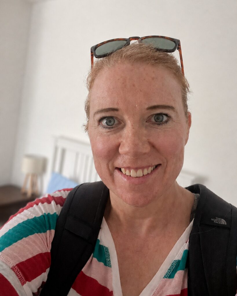
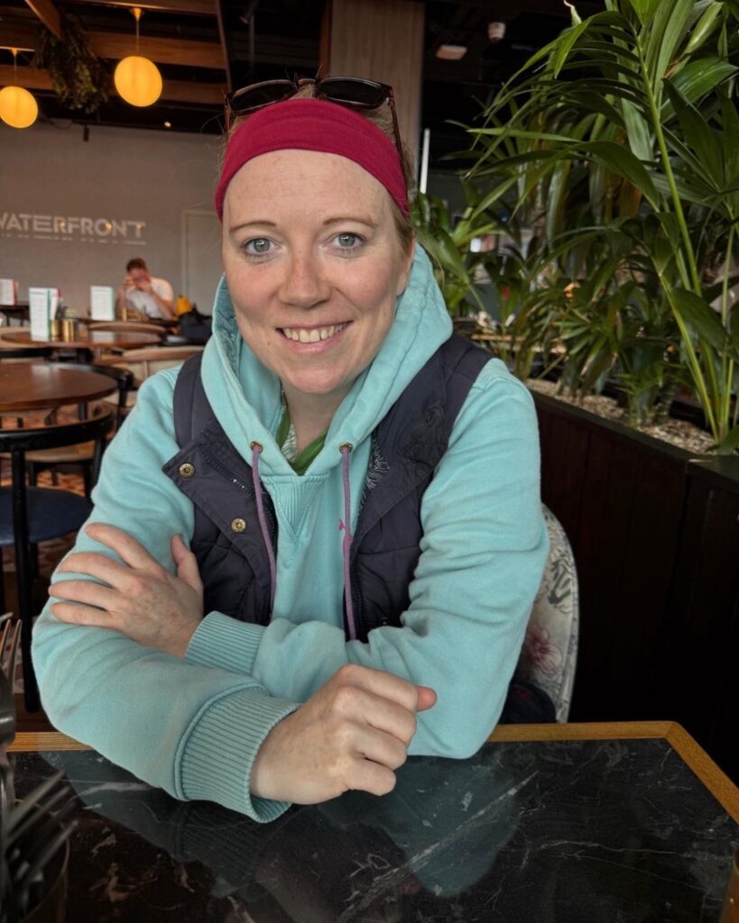
I initially had low cortisol and was on steroids but luckily this function has now recovered. I am back to work on a phased return and being well supported by my team and line manager. I am getting psychological support through work for what I have been through. I try and go swimming and socialise as much as I can, but I need to always listen to my body, pace myself, and be mindful of my fluid balance! I feel like I have a second chance at life and knowing the damage both a craniopharyngioma and its treatment can do, I have been exceptionally lucky and feel blessed.
“I feel like I have a second chance at life and knowing the damage both a craniopharyngioma and its treatment can do, I have been exceptionally lucky and feel blessed.”
I know that my journey is far from being over. I will need MRIs for the rest of my life. This type of tumour has a high chance of recurrence and if that happens I will need radiotherapy which could further damage my pituitary gland. At three months post-op, however, my MRI was completely clear. I’m trying to live life to the full. I won’t let the experience define me and will use it to live a better life for both myself and my family who went through this with me every step of the way.
I’m finding it’s helping to write a book about it all. Going from professional to patient in the NHS has been eye-opening in many ways and on many different levels and I feel I can better relate to my own patients now. My advice to others having any pituitary condition is to keep living life to the full, but listen to your body and trust your gut! And keep pressing the professionals for a proper diagnosis.

Have you found this helpful?
If so, please support us to bring useful information to more people affected by pituitary conditions.